The answer
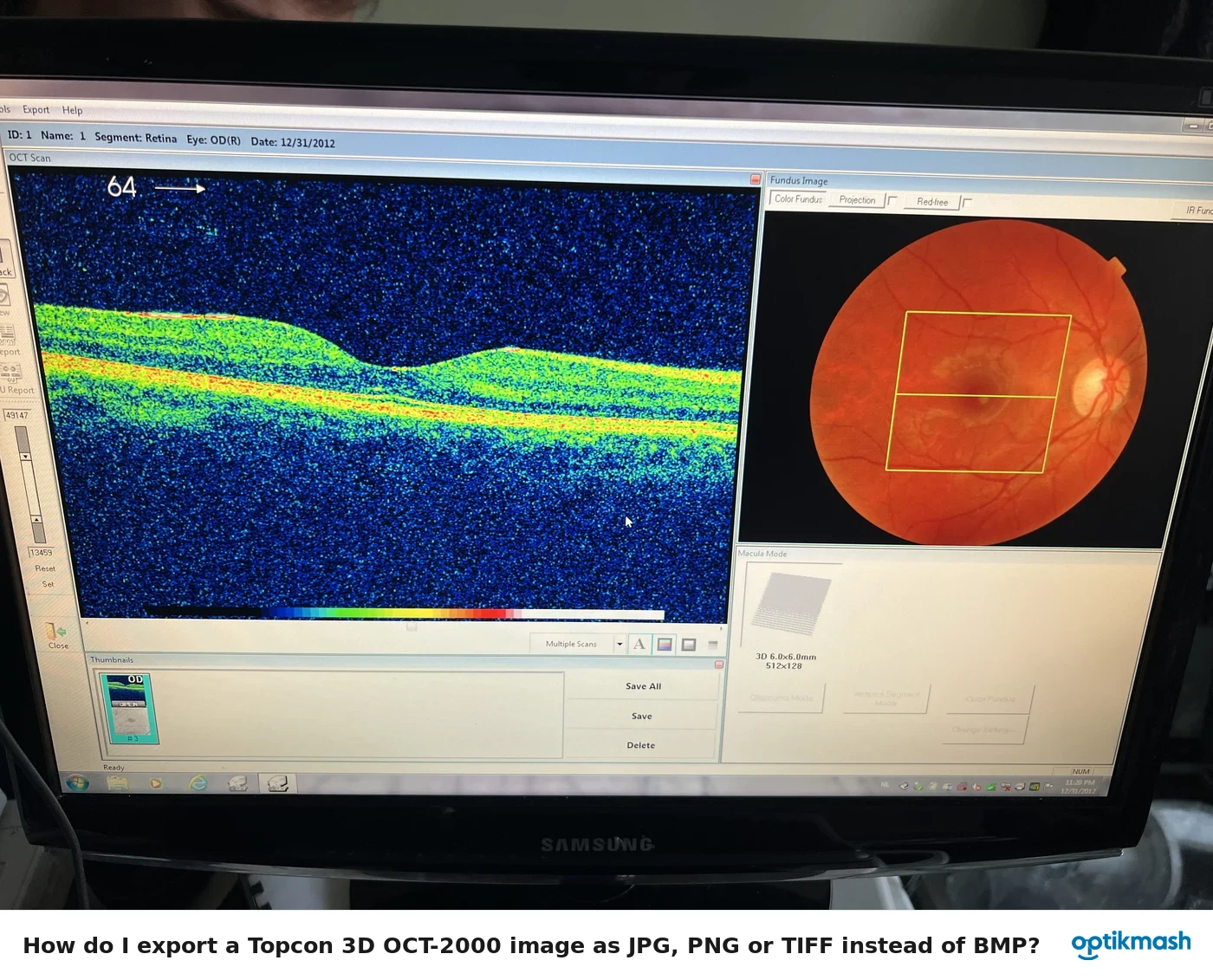
After the shadowgram is positioned with the color fundus image, double-click a point in the tomogram or tomogram display area. The Topcon 3D OCT-2000 then shows the corresponding location as a green line and cross mark on the fundus image, with thickness data in the Thickness graph area.
Filed under Image quality
Pin-point Registration™ links the 3D tomogram data to the color fundus image through the shadowgram. It is useful when the operator needs to inspect the tomogram at the exact location of a visible fundus feature.
- Ensure the shadowgram and color fundus image are correctly positioned.
- In the tomogram display area or auxiliary tomogram display area, double-click an optional point.
- Read the green line and cross mark in the fundus image display area.
- Use the linked tomogram position and the Thickness graph to inspect the corresponding data.
The center of the cross mark corresponds to the location of the white line on the tomogram. Double-clicking a point directly on the tomogram performs the same process.